
Foreign body in the stomach of dogs refers to a common disease in dogs that the dogs ingest indigestible foreign bodies, which cannot be excreted from the body, and stay in the stomach for a long time, causing various gastrointestinal diseases and gastric dysfunction.
Figure 1 Sick dog with hunched back and abdominal pain
1. Basic information of the case
Samoyed, three years old, weighing 30kg, male.
II. General and special inspections
1. Clinical examination The color of the conjunctiva and gums of the dog was flushed, and the body temperature was 38°C; the abdomen was palpated, and the patient showed restlessness, severe abdominal bulging and gag reflex.
2. Routine blood test
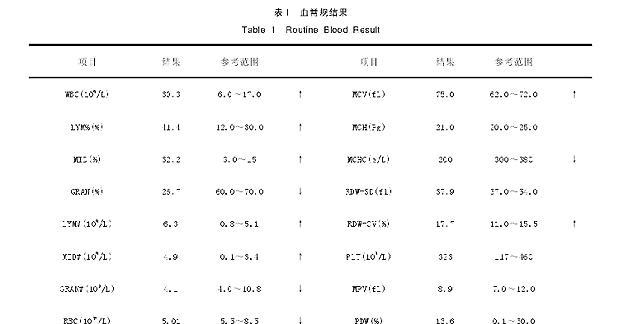
Analysis of blood routine results: white blood cells were higher, LYM% (%) and MID (%) were significantly increased, and red blood cells were decreased, indicating that the dog currently has an inflammatory reaction and infection.
3. Blood biochemical examination
Analysis of serum biochemical results: ALB was increased, AMYL was increased, and GLU was decreased, indicating that the current pancreatic metabolic function of the dog is impaired.
4. Imaging examination
Figure 2 X-ray inspection results
From the X-ray film, it can be observed that there are irregular projection areas with high density, which are suspected of foreign bodies in the stomach, and there are projection triangle areas with high density, which are suspected of foreign bodies in the stomach.
5. Observation results of B-type ultrasound instrument
Figure 3 B-ultrasound examination results
From the B-ultrasound examination results, it can be seen that there is a strong echo ring in the stomach, which is round, and some are accompanied by sound shadows.
Figure 4 Fasting infusion therapy
III. Surgical treatment
Extensive shaving and disinfection were performed on the front of the xiphoid cartilage of the dog's abdominal wall to the back of the umbilicus. The skin was first incised on the midline of the abdomen between the xiphoid cartilage and the umbilicus, the subcutaneous tissue was bluntly separated, and the abdominal wall muscles and peritoneum were incised.
Figure 5 Observation of incision recovery at each follow-up visit
IV. Experience and experience
1. Preoperative laboratory examination of animals is very necessary, so that the operator can better make a correct assessment of the operation; for dogs that have been diagnosed with gastric foreign bodies, especially those with large foreign bodies, maintenance
2. The number of foreign bodies removed by surgery should be compared with the results of imaging examinations. After surgery, X-ray and B-ultrasound should be performed on the dog to diagnose whether there are residual foreign bodies.
3. The general clinical manifestations of foreign bodies in the stomach of dogs are loss of appetite and vomiting, especially when feeding again after the onset.