
Chisha
1. Pathogenic analysis
1, Cinobacteria
New Cryptococcus Neoformans is a fungus that can infect dogs and cats. In contrast, dogs have less infections than cats. It is likely to enter the body from the respiratory tract, and some animals may still spread to other organs. In cats, clinical symptoms are mainly manifested in nasal cavity, central nervous system, eyes or skin and subcutaneous tissue infections. In dogs, the symptoms of central nervous system are the most obvious. Pulmonary infection is common in dogs and cats, but they rarely show corresponding clinical symptoms (such as cough, dyspnea, snot, etc.).
2. Empty mold
Aspergillus Fumigatus exists as a resident bacteria in many animals. But in some dogs and very few cats, it is a pathogen. These organic bodies can form fungal plaques (that is, "fungal cluster") that infringe the nasal mucosa. Generally speaking, animals are caused by some other nasal diseases, such as tumors, foreign bodies, trauma or immune defects, which leads to a reduction in the resistance of the animal body, and then secondary fungal infection. In those healthy animals, it is likely to be caused by excessive contact with shades. Another fungus-peniculia can cause symptoms similar to shaloma.
The pathogens that cause fungal rhinitis are mainly shaloma. Strains in different regions may be different, such as tobacco mold, green mold strain or spore strain. But its pathological foundation is destructive rhinitis. Although the mold is common in nature, it is mostly concentrated in some special environments, such as dry grass piles or cut grass with wet or storage conditions. What I still do not understand now is why some dogs actively infect and generate a large amount of mycelium and fungal colonies, although most dogs have the same amount of spores as normal as normal.
Of course, some susceptible factors are also implied in a small number of cases, such as trauma, sepsis infection, or other complications (including nasal tumors). The main cause of humans in western countries is severe immune disorders, but it has not been confirmed on dogs or cats. Cell immune injuries caused by cocoal mold may be caused by bacteria rather than animals.
2. Clinical features
Nasal molds can occur in dogs of any age and varieties, and are the most common in male puppies. Cats are rarely infected. Smooth may appear. Culminated purulent (blood or no blood) or blood -based nasal secretions. This nasal secretion may be unilateral or bilateral. Some may have sneezing. Penalized facial sensitivity or pigmentation and hair removal at the nostrils are the most representative manifestations of this disease. The lesion rarely accumulates the lungs.
Full -body molds are caused by Aspergillus Terreus or other types of molds. Unlike smoke mold, this systemic infection is often originally original in German shepherd, and it is usually fatal. This disease is not rare, and there are no reports of nasal symptoms of the diseased animals.
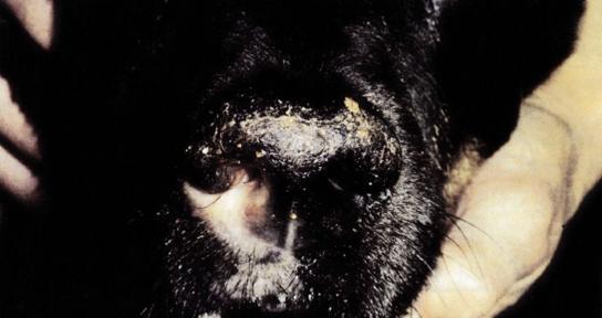
Figure 1: Sonic nostrils and mild ulcers occur
3, diagnosis
The discussion was first recorded in 1905. Significant molds were considered to be a cause of a dog producing chronic nasal secretions. However, it should be distinguished from other conditions before taking appropriate treatment. According to the results of nasal mirror examination and image diagnosis, it is found that infection has been confirmed during diagnosis. This may be due to the nasal secretion (main manifestations of symptoms). Before the pet saw the veterinarian, he was licked from the nostril or inhaled to the tail of the nasopharynx, and sometimes it may be swallowed directly.
Only a separate examination can not be diagnosed with mold disease. It is necessary to comprehensively evaluate the clinical symptoms performed by the dogs. In addition, epithelials are still a chance of pathogenic bacteria, so other potential nasal diseases must be considered. Bonococcal rhinitis of dogs should be identified with tumors, chronic rhinitis, nasal foreign bodies and dental diseases. Some more common causes may only consider whether they have direct evidence in the diagnosis room (such as cleft palate). By investigating its medical history, preliminary screening of different possibilities.
Investigation of the History of the disease
When the animal owner drives to see a doctor, the type of secretion and its unilateral or bilateral diagnosis will not help much. Because secretions can be transformed from early pulp types to mucus type or even direct nosebleeds. These secretions can be seen in many nasal diseases. Unilateral nasal secretions cannot be explained.
Facial pain is a characteristic. In some cases, animals may be manifested as spasm or even nasal bone collapse. Animals may also have non -nasal symptoms, such as decreased appetite and slow behavior.
Many animal symptoms do not show nasal secretions. People can check whether they have facial pain by rubbing their noses or gently patting the nose or anterior frontal bone. Its specific diseases are external nostril ulcers and decolorization (as shown in Figure 1), and there may be excessive skin keratosis. And it can be found that its respiratory tract airflow is normal, which helps to identify nasal tumors at the damage to the airflow.
In summary, dog fungi disease may be mainly manifested in: facial pain, exterior nostril decolorization or ulcers, and normally air flow.
Four, imaging examination
The X -ray signs of nasal mold disease are increased in the lesion area with a clear internal limit of the nasal cavity and the permeability of the ray permeability of the front end. The representative is that although the surface of the plow bone is uneven, the plow and facial bone have not been damaged. In the case of very few serious illnesses, these two parts and screens may occur. There may also be opaque liquid high -density shadows on X -ray. The liquid shadow in the frontal sinus is mainly caused by the accumulation of mucus caused by the infection or exclusion of the part. In some animals, only forehead infections are manifested. At the tail, a mixture can be found through X -ray photos on the nasal sieve part. In some special cases, granulation or fungal balls will appear.
Figure 2: X -rays show that there are obvious local nasal nasal cavity dissolved in the nasal cavity, and the plow bone is still complete
CT is the most effective image diagnostic method. When CT is required, there is no doubt that nuclear magnetic resonance imaging (MRI) is required. However, in practice, there are rarely one of the two ways of any way, and the specialty centers that shadow disease do not need to be mentioned for diagnosis and treatment are not required.
The morphological diagnostic characteristics of all rositum damage are similar. The damage to the loss obviously causes the space to be empty or the rays that look like they can pass through sexual cavities, and the height becomes thin (CT), and the empty signal (MRI). According to the degree of change, you can first see the nasal abdomen or head.
Nasal examination. After a whole body anesthesia, an image examination can be performed to check the nasal cavity. The application of endoscopy is like ear mikes, joint endoscopes, soft endoscopes and special nasal mirrors that rely on practicality. Regardless of the use of the instrument, the characteristics of the search are the lack of the l chenic bone and the existence of dry fungal plaques, and the mucus flash. Nasal mirrors can also observe the erosion of the nasal armor and the white to green plaque changes on the nasal mucosa. If these phenomena are not observed, molds cannot be ruled out immediately. The part of the suspected fungal mycelium can perform cytological examination and tissue cultivation (biopsy samples or nasal swabs), and then confirm the diagnosis.
Figure 3: The outer nostrils of dogs suffering from nasal and mold diseases are performed at a nasal mirror examination. You can see the lack of the nasal nails and brown -red granuloma and white fungal plaques.
Figure 4: The cytology pictures of the nasal tissue can be seen in the smoke mold wire
Eyloma can be observed by biopsy and conventional staining of the infected nasal mucosa. If you need to observe more fine lesions, you can also perform special dyeing. Disease animals usually show neutral, lymphocytic or mixed inflammation. Multi -sampling biopsy for this disease, because the disease of the mucosa is usually multi -stove. If a fungal disease is found in the nasal mucosa, the existence of infection can be proved.
Unless the sample is taken from visible fungal plaque, it is difficult to judge the results of fungal culture. There may also be certain fungi in the nasal cavity of healthy animals, so false negative results may occur. In other words, only when the training results are positive and show the corresponding clinical symptoms, the diagnosis can be diagnosed.
A positive results of serum antibodies can also explain the existence of infection. However, antibody positive can only indirectly prove the existence of infection, because molds are rarely induced to produce antibodies as part of the normal flora. As a result, false positive and false negative results may occur.
Five, Treatment
Over the years, people have tried a lot of treatment. This fact shows that fungus is difficult to remove in all animals treated. Of course, people will doubt whether animal lack of immune response to make it sustainable/infectious, or or fungi has resistance. Everyone knows that fungal infection is difficult to treat, and its systematic treatment index is not as wide as most other traditional drugs. Early treatment was mixed with lumper nail cutting and scraping, but the latter's cure rate showed poorly. Later, oral treatment did not continue, because the absorption of the digestive tract is low, and it is difficult to reaches the minimum inhibitory concentration of the nasal mucosa. The effectiveness of drugs in the nasal mucosa will be restrained because most fungi does not invade the mucous membrane or tissue related to the mucosa (such as the nasal mirrors). Therefore, in the past 20 years, it has focused on local medication. Research elements are drugs, administration methods and treatment time.
The most commonly used is club mimazole, which directly acts in the infection position. After one month of treatment, the nasal examination is performed to determine the treatment effect. If there are still fungi existence, secondary treatment is needed. The use of antifungal drugs should be detected at the same time as liver merit, because it may cause liver injury.
Figure 5: Cymezole soaking treatment of nasal fungal infection
6. Promotion
Prognosis depends on how to invade fungi. Only the empty cavity is the best to participate in the prognosis. Animal owner needs to realize that no matter what kind of treatment is used, it cannot be guaranteed to succeed at one treatment.