
Analysis of cat megaesophagus
Cat megaesophagus is a relatively rare disease. If it is hereditary or congenital, cats can develop the disease after weaning, and the acquired disease can occur at any age.
I. Definitions
1. Whole esophagus dilation and cessation of peristalsis in adult animals.
2. Adult primary megaesophagus refers to unexplained dilatation of the esophagus.
3. Adult acquired giant esophagus refers to esophageal dilatation with clear cause.
4. Adult megaesophagus often causes reflux in dogs but is rare in cats.
II. Etiology
1. The etiology of many cases is unclear, leading to congenital categories.
2. Many pathological conditions can cause or relate to esophageal dilatation.
3. Acquired (secondary) causes include neuromuscular diseases, immune regulation disorders, hormonal imbalances, poisoning and inflammation.
III. Clinical symptoms
1. Food and fluid regurgitation occurs, but the time period at which regurgitation occurs after eating varies widely.
2. Swallowing pain is seen in inflammation and hyperdistension, manifested by hypersalivation, repeated swallowing, and abnormal neck posture.
3. Bad breath occurs with the fermentation of food.
4. Shortness of breath (wet cough, rales, dyspnea) occurs when foreign body pneumonia occurs.
5. Malnutrition can lead to severe cachexia or weakness.
6. Symptoms related to secondary causes will be seen:
A. Muscle pain and polymyositis stiffness.
B. Neuromuscular weakness.
C. Gastrointestinal symptoms due to poisoning or Addison's disease (hypoadrenocorticism).
IV. Diagnosis
1. It is important to have an accurate medical history.
A. Confusion between vomiting and reflux can easily lead to misdiagnosis.
B. Giant esophagus is suspected in adult animals, especially dogs with a history of reflux.
2. Physical examination may provide clues to the cause of secondary giant esophagus.
A. Note muscle pain, weakness, neurological deficits, and changes in mental status.
B. Skin abnormalities can be seen in adrenal cortical dysfunction.
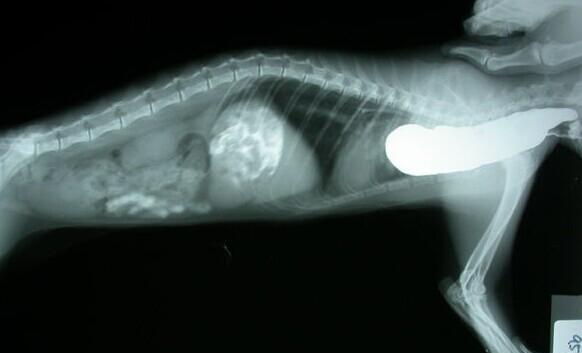
3. X-ray photography is appropriate.
A. General radiographic examination of the neck and chest can detect gas, fluid, and food accumulation in the esophagus.
B. Alveolar opacity can be seen in foreign body pneumonia.
C. Barium meal angiography of the esophagus can show its expansion and other structural abnormalities.
D. Fluoroscopy (if appropriate) can obtain useful information on giant esophageal retardation.
4. Laboratory diagnosis is beneficial.
A. The most basic laboratory tests include complete blood count, comprehensive biochemical test and urinalysis.
B. Myokinase analysis can determine myopathy or myositis, and serum cholesterol can be used to diagnose corticosteroid dysfunction.
C. All suspected myasthenia gravis and indeterminate cases of giant esophagus should be measured for acetylcholine receptor titers.
D. Clinically suspicious cases can be tested by traditional laboratory.
5. Esophagoscopy is generally not required for the diagnosis of giant esophagus unless tumor or other structural abnormalities are suspected.
V. Treatment
1. Animals with acquired giant esophagus can be treated at any time for the factors that cause esophageal dilatation, and most of them will improve the function of the organs.
2. Congenital giant esophagus and acquired giant esophagus ineffective drug treatment can be treated symptomatically.
A. Elevate the position of the feeding trough to facilitate the passage of food into the stomach.
B. Usually feed a small amount to meet physical needs.
C. Change the hardness of the food (compared with the bolus) to see which diet is best tolerated.
D. Animals with severe aspiration pneumonia require special attention.
3. After gastrography, feeding with a tube can not only ensure the supply of nutrients, but also reduce the risk of aspiration.
4. Broad-spectrum antibiotics can be used to treat bacterial infections.
5. The effect of function-promoting drugs has not been confirmed.
A. Cisapride (promoter) 0.5mg/kg, PO.BID can improve the esophageal function of cats.
B. It has been reported that it may improve the clinical symptoms of giant esophagus in some dogs.
6. Guardianship
1. Review every 1 to 2 months to monitor the development of the disease.
2. Repeat chest radiograph to confirm foreign body pneumonia with esophageal dilatation.
3. Adult megaesophagus generally has a poor prognosis.
A. Animals with primary disease often die or are euthanized due to disease.
B. Animal drug treatment of acquired diseases may be effective.
C. The prognosis of giant esophagus due to severe muscle weakness is good.