
The bone is also called the knee bone, which is a small bone under the tetraus muscle extension of the thigh femoral femur, which constitutes an joint with the fare of the femur. In front of a normal canine femur, there are quite deep grooves from two bone ridges, that is, the trench. The dislocation of the bone refers to the stretch of the crickets when the knee joint is extended, and it is suddenly dislocated from the ballast. It can be divided into internal dislocation or exogenous dislocation. It is more common in clinical disclosure. Divided into congenital and acquired nature. Congenital femoral bay deformities; the distal end of the femur and the near -end of the tibia are poor; the tibia is rotated, which makes the tibial roller move inward. The acquired nature is more common in trauma such as: jumping, collision, and strong pulling the quadriceps due to severe exercise or the internal and external ligament of the knee and the torn ligament in the knee. Clinically, bone dislocation can be divided into 4 levels according to the severity of the dislocation. The lightest man is that the bone is occasionally discharged and can be reset. The severe permanent dislocation can cause pain, cartilage injury, clamor of animal hind limbs, cartilage injuries, clamor , Even paralyzed.
The incidence of bone dislocation in all genetic diseases is 7.2 %, which is the highest among all congenital defects. It often occurs in small breed dogs, and large dogs also occur. The disease is one of the more common diseases of the teddy dog. The incidence of small bodies (such as toys, tea cups) is higher than that of other bodies (mini or larger) dogs. The condition continues to deteriorate and irreversible).
I. Case Introduction
Bichon, 9 months, female, 3kg, recently suddenly hanging the left back of the left limb, dare not bear the weight, no external injury history, a touch of pain without pain, and occasionally walking normal.
2. Diagnosis
Diagnosis methods are divided into consultation, visual diagnosis, palpation and X -ray examination. Ask the owner's onset time and process, observe the posture of the natural state of the back limbs, and then articulate the joints of the back limbs, observe the condition of the joint activity, and touch each part of the joints when stretching and flexion. When a dog is broken, the disease should be suspected of the disease through the painlessness of the entire affected limb. X -ray inspection, the positive film of the knee joint shows that the bone is located on the inside or outside of the femur instead of the femoral bay. At the same time, it can be seen that the trench becomes lighter, the tibia proximal bending, and the angle of the tibia joint.
Examination found that the backpage has no pain in the hind limbs, and both sides of the bones have different degrees of inner dislocation, and the left hind limbs are more severe (see Figure 1). The hand can be reset to the car ditch, and it will be removed immediately after loosening. X -ray inspection shows that the bone is located in the inside of the boat (see Figure 2). Diagnosis is dislocated on the inner side of the twin limb bone.
Figure 1 Bilateral stubborn stubborn inner dislocation
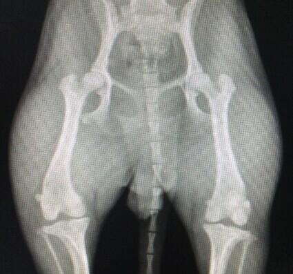
Figure 2 X-ray check shows the bone from the bone out of the bone
Three, Treatment
The mild inner dislocation of the bone can be overlapped through the outer side of the knee joint; the method of the treatment is invalid, the stubborn stubborn dislocation, shallow car ditch, or the short -to -boat cricket, etc., and generally deepen the correction of the knee joint trail; The exterior sides of the tibial bone is suitable for dislocation due to the tibial rotation.
The diseased dog adopted knee joint deepening surgery. The surgical path is the outer side of the knee joint, the incision begins with the near -near end of the trail, and is extended to the remote end of the travexed and ligament of the traveler and the outer end of the tibial bone (see Figure 3). Cut the skin, subcutaneous tissue, deep fascia and joint capsules in turn, showing the knee joint; the joint cartilage of the cutting of the car (see Figure 4), remove the cartilage wedge from the bone groove (see Figure 5), and then remove the bone from the incision, Deepen the tray (see Figure 6, Figure 7). Put the soft wedge back into the steamer (see Figure 8). After the bone is reset, the knee joint is flexed and extended to check its stability. If the bone is still in situ and the incision is conventional; if the bone is still off, the outer signs of tibial tibial can be performed.
Use superposition sutures to overlap the fascia connected to the outer fascia and the bone; the first layer uses a button suture to close the joint cavity to complete the 1st layer of suture (see Figure 9); then the second layer of suture is performed. The fascia on both sides of the suture (see Figure 10); clean up the wound, regularly suture subcutaneous tissue and skin; bandage the affected limb with a cushion bandage.
Four, postoperative care
After surgery, the bandage of the knee joint for 3 days (see Figure 11), limited joint exercise, avoid exercise within 1 week, and exercise moderately within 2 weeks; moderate exercise to promote the recovery of the affected limb after 2 weeks. Symptoms and anti -inflammatory inflammation after surgery.
After 1 week, the two weeks can stand on the limbs, remove the stroller in 10 days, and walk around after 2 weeks, but the gait is a bit stiff. After 1 month, you can walk normally.
Figure 3 Cut the skin on the front and outer side of the proximal bone
FIG
Figure 5 Removal cartilage wedge from the bone groove
Figure 6 Remove the bone from the incision to deepen the stepping
Figure 7 Baldling Sink deepen
Figure 8 joint cartilage deep into the femoral groin
Figure 9 button sewing closed joint cavity
Figure 10 The surface of the outer edge of the joint capsule and the surface of the inner edge
Figure 11 The bandage bandaged knee joint after surgery 11, limited joint movements, and walking exercise within 6 weeks